I got the Radiology report today on the recent MRI, everything is “normal”, no sign of disease.
I wonder why I have pain there, hmmm strange…
Hi Jon, yep, strange happens unfortunately. I do hope someone can come up with some answers for you soon!! On a good note, I guess no disease means there is hope of full recovery, which would be awesome!!!
1 Like
Well at least it might show that the Biologics have been helping.
The report needs to be challenged, as this could jeprodise my getting medications, and pain management of the area.
Ahhh… yes… soo true… Challenging a report might be difficult, perhaps your rhuemy might see things differently when he/she looks at the MRI pics. If you are finding benefit from the biologics hopefully docs will keep you on them. Here’s hoping!!! Cheers!!!
Hi Jon, is there anyway to get a second radiologist to look at it? I know if they don’t slice the MRI perfectly orthogonal to your spine it can look pretty strange and be hard to interpret, and I’m certainly no radiologist, but… that image looks a long way from symmetrical to me.
As you’ve had more experience with looking at them, I’m sure you can see even more spots than me that are to be questioned, in an ideal world if you could get your second opinion radiologist to talk you through it all, that would be magic 
1 Like
You are probably right, it is a little tricky to figure out the MRI’s. I am back at square one, unknown SI joint pain… But the biologic seems to be helping, so that it good…
1 Like
Hi Jon, I’m in a similar situation and feel your frustration. My Achilles’ tendon is swollen and irritated. Yet my MRI was normal. That was 6 months ago and it’s still bothering me, so I had an ultrasound of it on Thursday. I’m kind of expecting it to also be normal since the MRI was. Makes me feel like I’m crazy. Something is causing the swelling and pain! My rheumatologist diagnosed me with PsA despite the normal imaging. I also have SI Joint pain, and it can get really painful… I hope you get some answers. Did they inject you with dye? I’ve read that can help the radiologist view the soft tissues better. Maybe it’s a tendon issue and the dye would help? I did not get the dye but wish I had.
Yes, it was with contrast. No inflammation, so I would think that would mean the biologics are working.
I to had a lot of heel issues tied to enthesis inflammation.
2 Likes
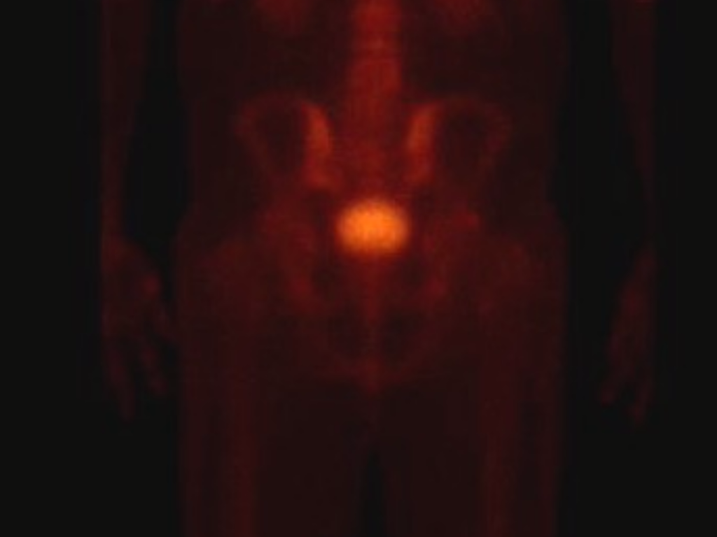
I think it is safe to say, I did have run away inflammation two years ago, before starting biologics.
Here is a nuclear bone scan, you can see I am lit up like a Christmas tree, the SIJ’s are fired up.
There was an interesting study done a couple of years on Remicade, showing that even for those (with significant erosions) who did not get better subjective scores (either Rheumy or patient), the erosions hasn’t progressed, so it is possible that the biologics is helping on the bone inflammation, even if it doesn’t feel that way.
What I meant from my first post though, was that to my completely untrained eye, it doesn’t look normal. Perhaps it is, or perhaps it is normal in that there is no active inflammation, and the erosions haven’t worsened (I imagine they would still have the potential to hurt if they are still present). Either way, I believe you have the right to ask the question and try to understand what the radiologist means by normal, and if the asymmetry is an imaging affect or an old erosion 
1 Like
That is encouraging about the biologic.
I understood what you mentioned, I think you might be right about the angled images. It would be nice to know that it is a MRI anomoly. I do know that my SIJ hurts like hell, and that Remicade is helping relieve some of the symptoms, fatigue is less, and I am more flexable, also muscles are more relaxed, I don’t know if that is caused by PsA, my previous doctors said it was Fibro. My mother has fibro, I touch a tender point and she jumped a foot off the chair!
The tech that did the ultrasound of me, laughed about the Fibro dx, he mentioned widespread enthesis envolvement as the posible cause, unofficially of course. He would show me the different images on ultrasound; enthesis, bone spurs, tears, etc. I think US imaging is the best way to monitor PsA, yet the Rheumy’s have yet to get on board with it. Next Rheumy, I will ask before the first appointment, “does the doctor use or have access to diagnostic muscular cellular ultrasound imaging?” This will be a deal breaker…
Hi there Jon_sparky, I read this today and thought of your post… I’m not 100% if it’s relevant or not, and you have possibly already seen it, but you might find it interesting.
" It is important to point out that a normal MRI scan, particularly in the spine, does not exclude enthesitis. This is because (a) bone oedema may be absent in the presence of enthesitis and (b) the enthesis being a relatively avascular structure does not readily accumulate fluid, hence is not well seen on MRI. Furthermore, the spatial resolution of MRI in the spine is quite low. Therefore abnormalities may not be appreciated. This means that in patients with inflammatory back pain the possibility of SpA cannot be excluded on the basis of a normal scan or normal blood investigations. It remains to be determined whether normal MRI scans are associated with less likelihood of future spinal fusion. For the same reason, MRI of synovial joints may not show enthesitis because of an absence of bone oedema at sites of enthesitis; moreover the soft tissue inflammatory changes associated with synovitis may mask enthesitis-related changes."
This is found on: http://www.arthritisresearchuk.org/health-professionals-and-students/reports/topical-reviews/topical-reviews-autumn-2009.aspx
In the paragraph headed “How can imaging be used to diagnose enthesitis?”
2 Likes
Thanks, it is helpful!
1 Like
I read up on the criteria for Dx of SPA using MRI, it is a joke, no wonder so few get diagnosed before 10 years. Erosions are disregardedfatty cysts are too, even the auther was lamenting how they needed to revise the criteria.
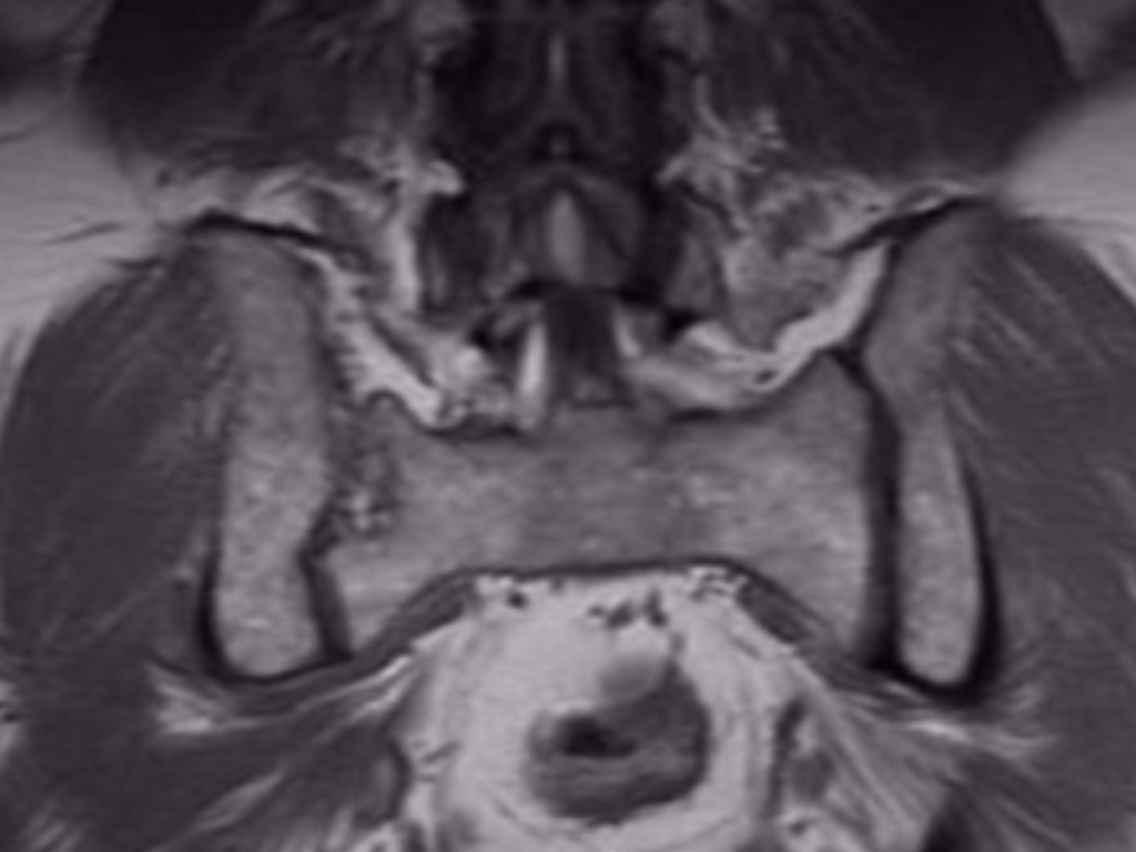
I came across this strange finding on my illiac bone, what do you guys think it might be? Looks like a bone wart…

Oh here is the criteria for Dx of PsA aith MRI.
http://www.elsevier.es/en-revista-radiologia-english-edition--419-articulo-new-asas-criteria-for-diagnosis-S2173510714000056?redirectNew=true
Jon, this is an area I really should learn more about (I don’t even know which one is the iliac bone!). Kudos to you for trying to understand it and taking control .

So, the illiac is the big flat bones that make up the hip, the sacrum is the triangle bone that supports the weight og the torso and distributes it to the legs.
The mri imageis a angled slice from the back, slice through the illiac bone, you can see the lumbar spine starting the the upper part of the image, the little triangle is the Sacrum.

Here are some of the findings of my “normal” SIJ. This is with contrast.
I think the bump is from the bone marrow biopsy, I had 2 years ago! It was funny at the time, I was in twilight sleep, and they were hammering away took about 8 swings with a mallet! I was laughing in my mind…