My experience of imaging is that there’s usually very little feedback so I’ve never got to grips with it. But I think what you’re saying is that imaging is only as good as the people who interpret it(?) So far it really has been all about disease control and obviously I appreciate that but I’m living with a body that’s got more complex. I think it’s time to know more and it’s great that my rheumy has acknowledged my concerns.
Funny you should mention about the BME bone marrow edema, I was going to start a tread about that! I was reading about bone inflammation, when researching the x-rays on my hand, I came across references to BME, and remembered that I questioned my spinal surgeon about the BME in my vertebreas (all), and the article touched on it regarding AS, that this may be a early sign that the area will ankylosis or fuse later. Here is a CT image of my spine, notice the honey comb appearance, this is thought to be caused by bone inflammation.
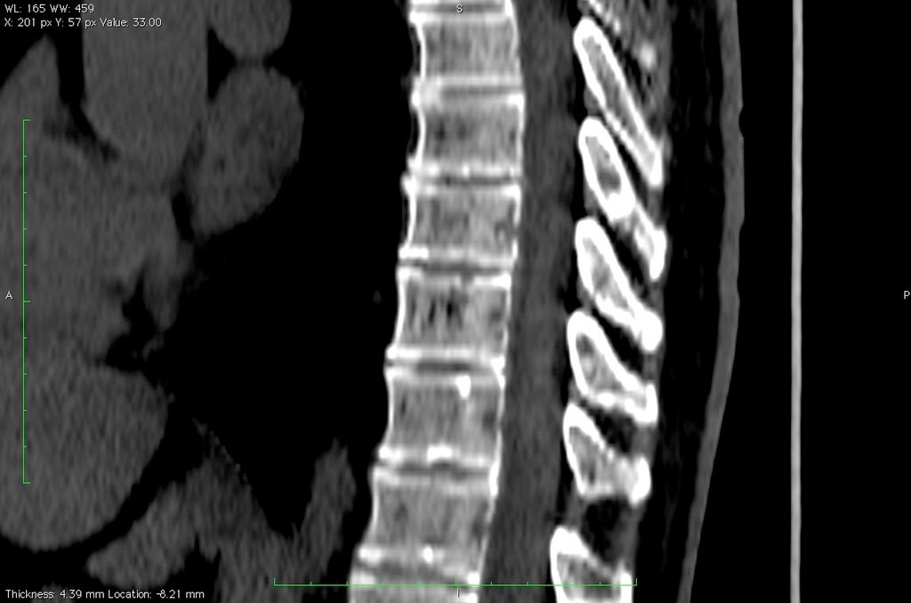
The area where the vertebrae touches the disc is a enthesis, and effected by PsA and AS, you can see the jagged appearance of it on my CT, from erosions.
This is a great discussion, and thanks so much Jon for posting your CT scan. Sometimes when a report comes back saying normal, and virtually no other comment, it’s good to have an idea of what ‘not normal’ looks like!
Or even when the gist is ‘not normal’ but virtually no other comment and at best you get to peer at the doc’s screen for a few seconds. A learning curve and no mistake! Thanks Jon_sparky!